


In vivo Fiber Photometry
Fiber photometry is a powerful tool used for detecting and quantifying bulk fluorescence in the brains of freely moving animals. This technique can be combined with targeted viruses that express activity-dependent sensors. The Centanni lab uses fiber photometry in combination with a number of novel calcium, glutamate, GABA, and other fluorescent sensors.

Viral sensor expression and fiber implant, with an example fluorescence trace aligned to behavior.
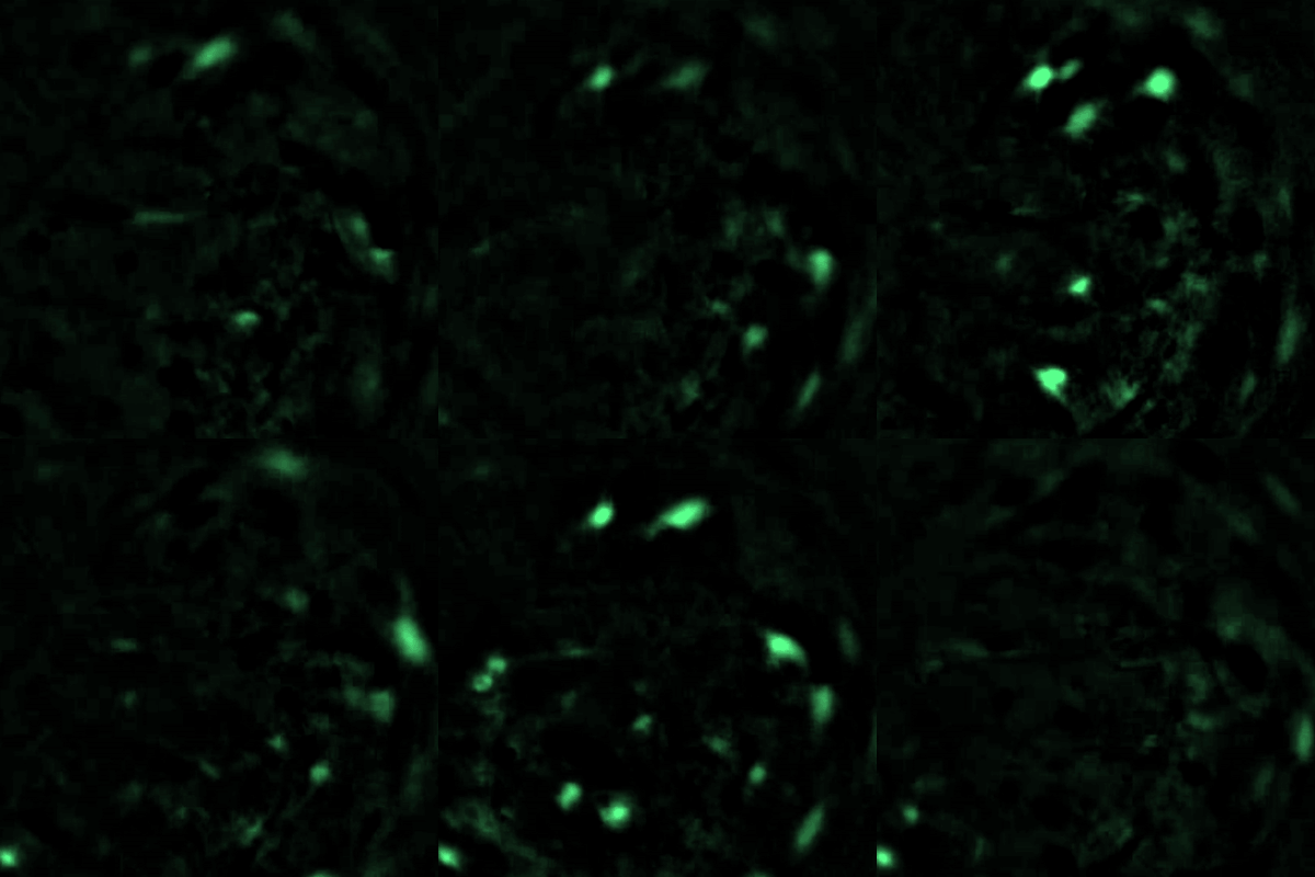
Single-cell calcium activity recorded with a head-mounted miniscope (shown at 2x speed).
In vivo Calcium Imaging with Miniscopes
Miniature head-mounted microscopes (Inscopix) allow us to record the activity of dozens to hundreds of individual neurons in freely moving mice. A small lens implanted above a brain region of interest relays fluorescence from a genetically encoded calcium sensor (e.g., GCaMP) to a lightweight camera on the head. Unlike fiber photometry, which measures the combined signal of a population of cells, miniscope imaging resolves single neurons. This lets the lab track how specific cells respond during stress, alcohol drinking, and other behaviors, and how those responses change across days and weeks.
Light Sheet Microscopy
Light sheet microscopy is an emerging technique that allows for imaging of whole delipidized rodent brains and other organs that can then be reconstructed to provide a single 3D image. Light sheet microscopy works by taking very small serial images by passing “sheets” of light through the tissue. This approach provides high resolution and near seamless reconstruction of specimens. The Centanni lab uses light sheet microscopy in combination with endogenous virus expression and immunohistochemistry to visualize whole brain circuitry with unprecedented spatial resolution.

Whole brain light sheet reconstruction showing two fluorescently labeled cell populations.
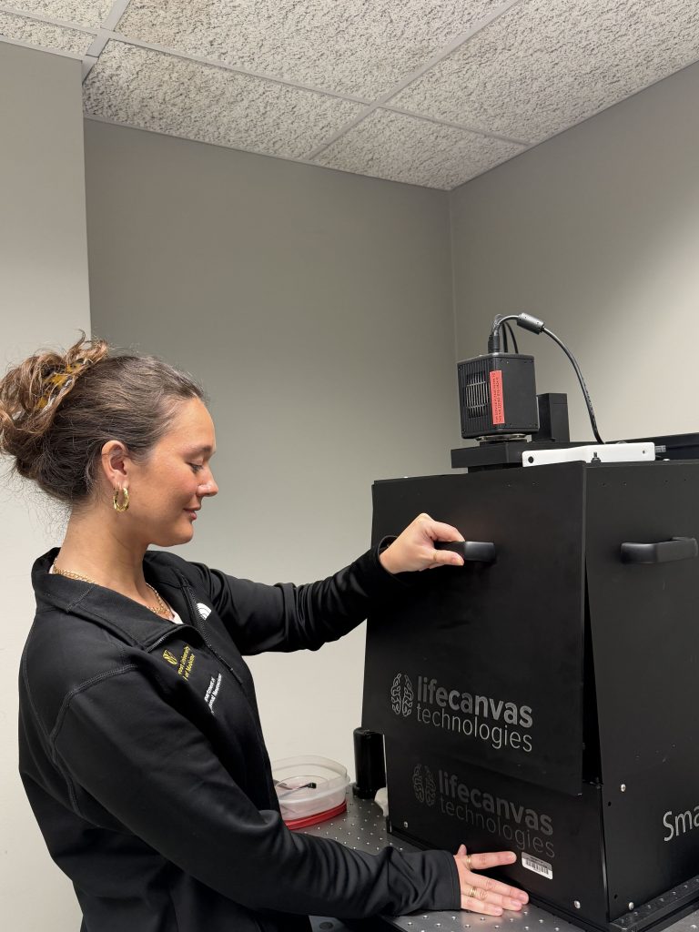
The lab’s light sheet microscope in use.
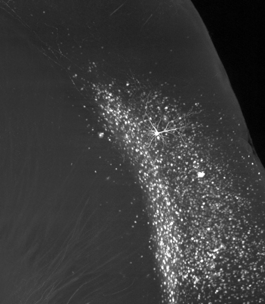
FosTRAP-labeled stress-activated neurons in the insula.
Activity-dependent Ensemble Labeling (FosTRAP)
Only a small fraction of neurons in a brain region respond to any given experience. To isolate these ensembles, the lab uses FosTRAP, a genetic strategy in which neurons that switch on the activity-dependent gene Fos within a defined time window are permanently labeled after a single dose of 4-hydroxytamoxifen. This allows us to tag the specific cells recruited by a stressor, then return weeks later to map their connections with light sheet microscopy, record or manipulate their activity, and compare them to neighboring cells that were not activated.
Translating Ribosome Affinity Purification (TRAP-seq)
To ask what makes these cells molecularly distinct, the lab uses TRAP-seq. A Cre-dependent, GFP-tagged ribosomal protein (eGFP-L10a) is expressed in a defined population of neurons, either cells that project to a specific target, such as insula neurons that project to the BNST, or a stress-activated ensemble captured with FosTRAP. Tagged ribosomes are then pulled down and the mRNAs they are actively translating are sequenced, revealing how stress and alcohol reshape gene expression in that population. Top candidate genes can then be knocked down in the same cells with Cre-dependent shRNA to test how they shape stress responses, drinking, and affective behavior.
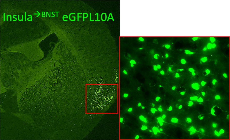
Insula neurons projecting to the BNST expressing the TRAP-seq ribosomal tag eGFP-L10a.


Patch pipette on a neuron in an acute brain slice, with a schematic of the recording.
Acute Slice Electrophysiology
Whole cell electrophysiology is an established technique optimized for probing individual neurons and recording electrical activity. Acute brain slices are maintained using artificial cerebrospinal fluid, and a micropipette is used to gain access to the cells. Once accessed, electrical activity is measured using a digitizer and amplifier. The Centanni lab uses this technique to characterize spontaneous and evoked activity of specific cell types in specific brain regions under specific experimental conditions (e.g., after alcohol exposure or stress).
Channelrhodopsin Assisted Circuit Mapping
The lab also uses a sub-technique called channelrhodopsin assisted circuit mapping (CRACM). This involves injecting a virus expressing channelrhodopsin into specific upstream brain regions to gain control of a circuit. A neuron in the target region is recorded from, and optical stimulation of the terminals expressing channelrhodopsin can help determine whether two brain regions are functionally connected.

Light flashes on channelrhodopsin-expressing terminals drive firing in a connected downstream neuron.
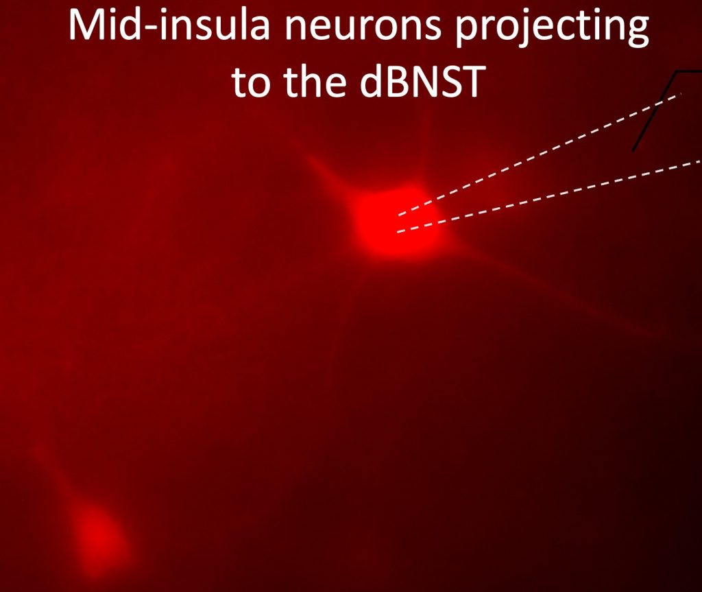
A mid-insula neuron projecting to the dorsal BNST, targeted for whole-cell recording.

Fluorescent in situ hybridization for Crh (red) and Fos (green), with arrows marking Fos-positive cells.
Fluorescent in situ Hybridization and Immunohistochemistry
FISH uses fluorescently labeled probes to “tag” RNA expression in cells. Immunohistochemistry is a technique that uses antibodies to visualize protein expression. Both techniques are widely used in the lab. For example, the immediate early gene c-fos, a proxy for neuronal activity, can be assessed in brain slices or whole brains to determine which cells are active under specific conditions (e.g., after alcohol exposure or stress).
ECG Biopotential Recording and Heart Rate Variability
The insular cortex helps the brain sense and regulate the body’s internal state, including the heart. To study this brain and heart connection, the Centanni lab records electrocardiogram (ECG) biopotentials in freely moving mice. From these recordings we measure heart rate and heart rate variability (HRV), the beat to beat fluctuation in the timing between heartbeats, which reflects the balance of sympathetic and parasympathetic control of the heart. Combined with fiber photometry and chemogenetics, this approach allows us to record insula activity and cardiac function at the same time and to test how stress-sensitive insula circuits shape the body’s autonomic response to stress and excessive alcohol consumption. Because HRV can be measured with the same logic in mice and in people, it offers a non-invasive bridge between our preclinical findings and future clinical biomarkers.
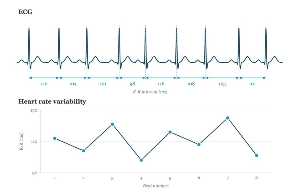
Beat to beat variation in the R-R interval, the basis of heart rate variability.

Chemogenetic and optogenetic workflows, from viral delivery to receptor activation and behavior.
Chemogenetics and Optogenetics
Chemogenetics and optogenetics use modified receptors or ion channels to gain control of specific circuits and cell types using either pharmacology (e.g., DREADDs) or light activation (e.g., channelrhodopsin) to allow for bidirectional control of neuronal activity. This strategy is used in the lab with our stress and alcohol models, and can be combined with a number of the other techniques highlighted here.
Preclinical Models of Affective Behavior
The lab performs several mouse behavior models to assess stress and negative affect, including the novelty suppressed feeding test (NSFT), open field test (OFT), light/dark test (LD), elevated plus and zero mazes (EPM, EZM), and startle response. The lab has adopted machine learning based algorithms to measure discrete mouse behavior that occurs during these tasks. DeepLabCut provides highly precise, unbiased, accurate measurements that can be used to categorize behavioral phenotypes that are specific to a condition (e.g., alcohol exposure or stress).

Overhead video from a behavioral test, analyzed with DeepLabCut pose tracking.